Friday 15 May 2015

SYED IRFAN ABID BUKHARI     03336366260      spl-education.blogspot.com
Guide to Tympanometry

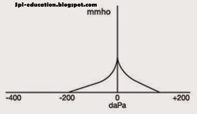
Tympanometry is routinely performed as part of our Full Diagnostic Hearing Test and is used mainly as a measure of middle ear function. This is achieved by changing the air pressure in the ear canal. Doing so changes the position and compliance (flexibility) of the eardrum and consequently the efficiency with which sound is admitted into the middle ear.
This is recorded objectively and the one is graphed against the other. Compliance and vibration of the eardrum and the admission of sound into the middle ear are greatest when the air pressure is the same on both sides of the eardrum. Thus, when Eustachian tube function is normal the peak in the graph (maximum compliance) is at normal atmospheric pressure (0 daPa).
The maximum compliance, and the pressure at which it is achieved, provides information about the middle ear and its contribution to hearing loss. Additional information about the middle ear and also about the inner ear can be obtained by using a loud noise to trigger reflex contraction of the middle ear muscles and transiently alter the maximum compliance.

Tympanometry provides information regarding the nature of the hearing loss, particularly the difference between a sensorineural and a conductive hearing loss. 
Type ATympanogram

Suggestive of normal middle ear function.
The peak of the tympanogram must be within +/- 100 daPa with a compliance of no more than 1.4 millimho (mmho) or no less than 0.3 mmho.
Type As (A shallow) Tympanogram
Suggestive of a less compliant middle ear system.
Patients that present with Type As tympanograms normally have a history of middle ear problems ranging from childhood otitis media, otosclerosis and tympanosclerosis.
The peak of the tympanogram must be within +/- 100 daPa with a compliance of less than 0.3 mmho (ASHA, 1990).
Type Ad (A deep) Tympanogram
Suggestive of a highly compliant middle ear system.
This may present in patients who have had multiple perforations in the past, or multiple sets of grommets. Patients with his tympanogram may also have an ossicular chain disruption.
The peak of the tympanogram must be within +/- 100 daPa with a compliance of greater than 1.4 mmho (ASHA, 1990).
Type B Low Tympanogram
Suggestive of middle ear dysfunction.
Patients with Type B low tympanograms can have otitis media. This is particularly common when a patient has a cold and is especially prevalent in children. Hearing loss may be present in cases of middle ear effusion. 
There is no identifiable peak with an ear canal volume within normal limits (between 0.6 – 1.5 cm3).
Management of Type B low tympanograms involves a tympanometry recheck in three months time to monitor whether or not the middle ear issue has spontaneously resolved. If this is has not occurred, an ENT referral is recommended.
Type B High Tympanogram
Suggestive of a grommet or perforation.
Perforations can cause a drop in low-pitched hearing and can progressively get worse. The most common causes of perforation arise from ear infections, trauma and extreme pressure changes i.e. diving and flying.
There is no identifiable peak with an ear canal volume greater than 1.5 cm3.
ENT referrals are recommended in almost all cases. It is important to note that an ear with a perforation is no longer waterproof and a swimming plug may be necessary in these cases. The presence of a patent grommet also often creates slightly elevated low-pitch thresholds.
Type C Tympanogram
Suggestive of Eustachian tube dysfunction.
The peak of tympanogram occurs at an admittance value more negative than –100 daPa (ASHA, 1990).
These tympanograms usually occur after a patient has had a cold or is about to get one. Patients with a long history of Type C tympanograms are at risk of cholesteatomas. Patients with Type C tympanograms are monitored every three months, or yearly if the patient has a long history.